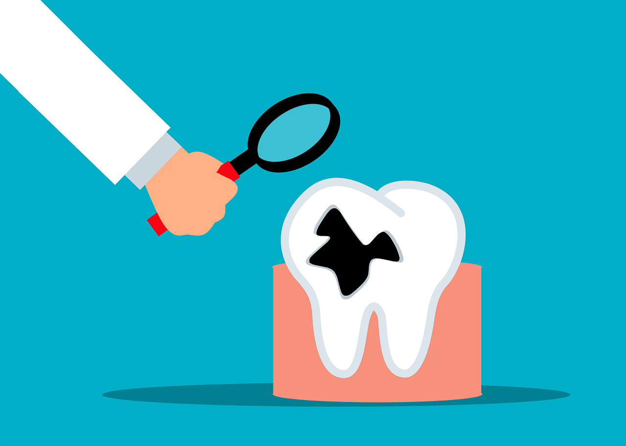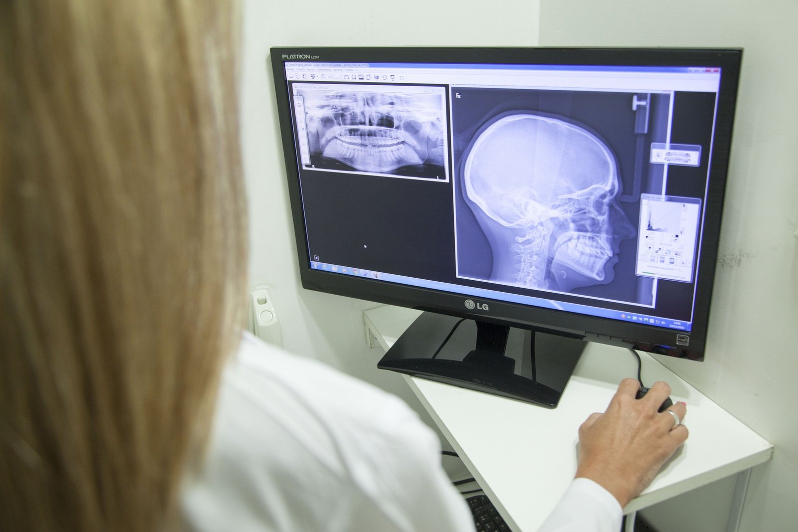
Dental radiographs play a crucial role in the diagnosis of a number of oral diseases, such as dental caries, periodontal lesions, and cysts and tumors of the oral and maxillofacial region. Dental radiography is referred to as “traditional” if it uses conventional X-ray films to expose diagnostic images. Among the intraoral radiographs are periapical, bitewing, and occlusal images, while a few extraoral images include panoramic and lateral cephalograms.
Intraoral radiographic examination is the cornerstone of diagnosis and treatment planning for any general dentist. The tooth and its surrounding structures are visible on the intraoral periapical radiographs. It assists with locating apical inflammation/infection, apical cysts, or any tumors, evaluating periodontal health, evaluating the condition of the tooth and alveolar bone after trauma, evaluating root morphology prior to extraction, and evaluating the placement and outlook for implants.
In addition to intraoral radiographs, the oral and maxillofacial region is often imaged with extraoral radiographs. They are used to diagnose trismus (restricted mouth opening), examine the size of large lesions, assess skeletal growth and development, determine the location of impacted teeth, examine the temporomandibular joint, and assess bone trauma.
To close the gap, recent developments in digital radiography are anticipated. Oral and maxillofacial radiology underwent a period of change from the traditional 2-D radiography to 3-D radiography, which lessened the difficulty of diagnosing the more typical lesion.
Dental X-rays can be taken conventionally (on film) or digitally (with digital sensors and a computer). Comparing digital dental X-rays to conventional dental X-ray machines, the radiation used is reduced by 80% to 90%.
Even though having a radiographic scan exposes you to some radiation, radiation is present all around us in nature and our surroundings, so it is impossible to avoid any type of radiation exposure. The average American receives 3 millisieverts of radiation annually from background radiation sources, according to estimates. Of course, the more dental x-rays you have, the more radiation your body is exposed to. It’s important to only get dental x-rays when absolutely necessary because excessive radiation exposure can increase your risk of developing certain types of cancer.
The radiation dose from a standard dental x-ray is about 10 micro Sieverts. More radiation is required and the amount could reach as high as 22 micro Sieverts if the dentist is using an older-style dental film. The radiation dose drops dramatically to between 2 and 5 micro Sieverts if the dentist uses a newer style of x-ray unit with a rectangular end rather than a round end. You will receive an exposure to radiation of about 180 micro sieverts if you undergo a full mouth series of dental x-rays (typically 18 x-rays).
The patient is exposed to radiation in the range of 9 to 26 micro Sieverts during a panoramic dental x-ray or OPG. You would receive about 50–60 micro Sieverts of radiation if you visited the dentist and had four standard dental x-rays taken in addition to a panoramic x-ray. This is comparable to the radiation exposure from spending six days on this planet Earth.
Many young people who receive braces or orthodontic treatment also undergo lateral cephalometric (or lateral ceph) x-rays. The radiation dose from a lateral cephalometric x-ray is approximately 5 micro Sieverts, which is equal to one half day’s worth of background radiation exposure.
You can see the jaw bones in three dimensions using Cone Beam Computed Tomography (CBCT) scans. This helps dentists diagnose fractures and oral pathology and can also help with the assessment of patients for dental implant placement. CBCT units can emit anywhere between 20 and 600 micro Sieverts of radiation, or 3 to 75 days’ worth of background radiation, depending on the brand.
The amount of energy that is absorbed when something or someone is exposed to X-rays is measured by the radiation dose. This is crucial because it is this energy absorption that can harm a person. To express dose, various units of measurement are used. Also, Diagnostic Reference Levels (DRL) are frequently used to express measurable doses from radiological procedures.
For intraoral radiography, the kerma-area product ranges from 26 to 87 mGy.cm2 and the entrance surface kerma ranges from 0.65 to 3.7 mGy while for panoramic radiography, the entrance surface dose should be between 3.3 and 4.2 mGy and the kerma-area product should be between 84 and 120 mGy.cm2. The kerma-area product for lateral cephalometric radiography ranges from 41 to 146 mGy.cm2 for adults and 25 to 121 mGy.cm2 for children.
In addition to the aforementioned, the effective doses for various radiographs vary, for example, intraoral radiographs are 1-8 Sv, panoramic scans are 4-30 Sv, cephalometric radiographs are 2-3 Sv, and CBCT scans range from 50 Sv or less for small- or medium-sized scanning volumes to mymedic.es 100 Sv for large volumes.
Less than a day’s worth of background radiation are typically emitted during intraoral and cephalometric dental radiological procedures. Even at the high end of the range, doses for panoramic procedures are comparable to a few days’ worth of background radiation that is similar to that of a chest radiograph. Doses for panoramic procedures are more variable. CBCT doses are extremely variable, but depending on the technique they may be tens or even hundreds of Sv of effective dose higher than traditional radiographic techniques. The usual dose ranges will probably change as a result of the CBCT equipment’s rapid technological advancements.
With the understanding of all radiation doses during x-ray exposure, it is now known that there is always a risk with x-ray exposure, but it is very low, and we should never be afraid and always go for a radiographic scan to help the dentists more accurately diagnose the pathology in the mouth and give you the best of treatment.